
2D, 3D, and 4D ultrasound all use sound waves to obtain information about a pregnancy, but they present that information in different ways. 2D is the basis of many clinical evaluations; 3D reconstructs a volume, and 4D shows that reconstruction in motion. No format is automatically “better”: the choice depends on the purpose of the study, gestational age, fetal position, viewing conditions, and the clinical questions that need to be answered.
This content does not diagnose conditions or replace a consultation, an emergency evaluation, or instructions from your healthcare professional.
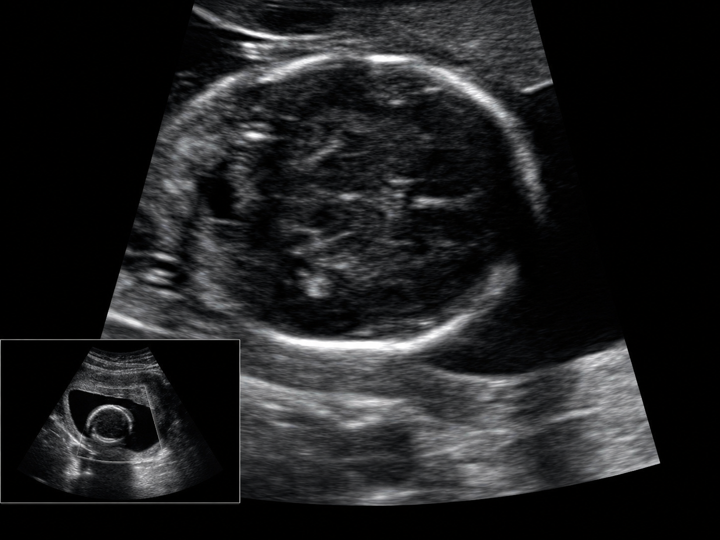
Differences between 2D, 3D, and 4D ultrasound
All three formats rely on the same ultrasound technology. What changes is how the information is acquired, reconstructed, or displayed. This comparison summarizes their usual roles without treating one format as a substitute for another.
| Format | How it looks | What it usually adds |
|---|---|---|
| 2D | Flat sections, usually in grayscale and viewed in real time. | It is the basis for evaluating anatomy, growth, cardiac activity, movement, placenta, and amniotic fluid, as indicated. |
| 3D | A three-dimensional reconstruction created from a volume of information. | It may provide a surface or spatial view and complement selected cases when the professional believes it adds useful information. |
| 4D | A continuous sequence of 3D images through time. | It may show movement, position, and some gestures when gestational age and viewing conditions allow. |
What a 2D ultrasound shows
A 2D ultrasound shows sections through the fetal body. Although some images may look abstract to an untrained viewer, these planes allow the professional to take measurements and systematically examine internal structures.
Depending on the study and stage of pregnancy, it may provide information about anatomy and development, growth, cardiac activity, movement, fetal position, placenta, amniotic fluid, and multiple pregnancies. The evaluation is not based on a single picture: it combines multiple planes, measurements, and gestational context.
The type and extent of the ultrasound should answer a clinical question. An image that looks less recognizable may therefore contain essential medical information that a surface view cannot provide.
What 3D ultrasound adds
3D ultrasound gathers information from a volume and presents it as a three-dimensional reconstruction. When fetal position is favorable, it may show surfaces such as the face, hands, or feet in a way that is easier for the family to recognize.
An easier-to-recognize image does not mean the study is more complete. 2D planes continue to provide essential internal information. In selected situations, 3D reconstruction may help the professional understand a spatial relationship or complement an observation, but it is not a universal replacement for conventional evaluation.
What 4D means and what you may see
4D ultrasound adds time, showing successive 3D reconstructions in motion. Depending on the stage of pregnancy and visualization, it may show hand or foot movements, changes in position, and some facial or mouth gestures.
These images may have emotional value for a family, but their clarity alone does not determine the baby’s health. They also do not replace the report or professional interpretation. The clinical purpose of the study should remain more important than obtaining an attractive image.
Why the face may not appear clearly
Ultrasound does not work like a camera. The quality of a surface view can change because of fetal position, hands or arms in front of the face, proximity to the placenta or uterine wall, the available angle, and other technical or pregnancy-specific conditions.
The professional may try different angles or spend time completing the necessary views. Even so, a clear facial image cannot be guaranteed at a particular appointment. Not seeing the face as the family expected does not by itself indicate a problem or invalidate the clinical information that was obtained.
CEMAFE does not guarantee a particular facial image. The priority is obtaining the indicated clinical information prudently and professionally.
Does 3D or 4D replace the clinical 2D evaluation?
No. 3D and 4D are complementary tools. 2D evaluation remains fundamental for examining internal structures, taking measurements, and answering many clinical questions during pregnancy.
Clinical organizations and health authorities recommend that ultrasound be performed by trained professionals, for a medical indication, and for the time needed. If family images are obtained, the prudent approach is to do so during a medically indicated examination without extending exposure solely to obtain a particular view.
What to expect during the study
The professional commonly begins with 2D images to guide the evaluation. When the clinical question, equipment, and conditions allow, 3D or 4D views may be added. The exact sequence is not the same for every patient and may change during the study.
Visual quality also varies from one appointment to another. A sharply defined 3D image is not the same as a complete study, and a less-defined view of the face does not mean the clinical information is insufficient. At the end, ask what was evaluated, whether the required views were completed, and who will interpret the report.
Useful questions before you leave
These questions can help separate clinical value from visual format and clarify next steps without trying to interpret the images on your own.
- What was the main purpose of the study?
- Were the necessary views completed?
- Did the 3D or 4D images add complementary information?
- Who should interpret the report?
- Do I need a follow-up evaluation?
- How can I keep and share the complete study?
Keep more than a screenshot
A screenshot or photograph of the monitor does not contain the complete study and may lose resolution or context. For future consultation, keep both the report and the available images or files, and share them with the professional overseeing the pregnancy.
At CEMAFE in Santiago de los Caballeros, you can ask about digital delivery through WhatsApp and downloading full-resolution images. Delivery makes the study easier to keep and share, but it does not replace the report or a conversation with your obstetrician or specialist.
To coordinate an obstetric ultrasound or ask about the 3D and 4D options available for your indication, contact CEMAFE through WhatsApp.
Frequently asked questions
Quick answers about this topic
Is 4D ultrasound better than 2D?
No. They are formats with different roles. 2D is essential for many clinical evaluations, while 4D may add a moving three-dimensional view when appropriate.
Can 3D or 4D ultrasound detect every condition?
No. Every ultrasound has limitations, and its findings depend on gestational age, fetal position, visualization, and the clinical question. It must be interpreted with medical history and other evaluations.
Can a clear image of the face be guaranteed?
No. Fetal position, angle, and other conditions may prevent a clear view. This does not by itself mean there is a problem.
Sources consulted
References used to prepare this guide. The content has been adapted into patient-friendly educational language.